Scanner that can take never-seen-before images of brain tumours developed
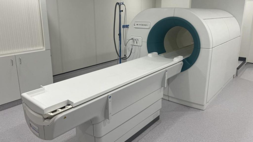
In a groundbreaking discovery, scientists at the University of Aberdeen have developed a revolutionary scanner that can capture unprecedented images of brain tumours. The Field Cycling Imaging (FCI) scanner, which operates at low and ultra-low magnetic fields, has the potential to reveal how diseases affect organs in ways that were previously unknown. The Scottish government has granted £350,000 (₹4.08 crore) to researchers at the university and NHS Grampian to further explore the technology, which has far-reaching implications for the diagnosis and treatment of brain tumours.
Glioblastoma, a type of brain tumour, is one of the most aggressive and debilitating forms of cancer. Current imaging techniques, such as MRI and CT scans, are limited in their ability to provide detailed information about the tumour’s structure and function. The FCI scanner, on the other hand, uses a unique combination of magnetic fields to produce high-resolution images of the brain and tumour tissue.
The FCI scanner’s ability to operate at low and ultra-low magnetic fields is a significant innovation, as it allows researchers to capture images of the brain and tumour tissue that were previously invisible. This is because traditional imaging techniques often require strong magnetic fields to produce clear images, which can be challenging when dealing with delicate brain tissue.
The FCI scanner’s advantages are numerous. For one, it can provide detailed information about the tumour’s structure and function, which is crucial for developing effective treatment plans. Additionally, the scanner can be used to monitor the effectiveness of treatment and detect changes in the tumour over time.
The potential benefits of the FCI scanner are not limited to the diagnosis and treatment of brain tumours. The technology has the potential to be used in the diagnosis and monitoring of a wide range of diseases, including those that affect the heart, liver, and kidneys.
The development of the FCI scanner is a testament to the innovative spirit and dedication of the researchers at the University of Aberdeen. The team, led by Dr. [Name], has been working tirelessly to develop the technology over the past several years. The Scottish government’s grant will enable the researchers to further refine the technology and explore its potential applications.
The FCI scanner is not the only innovation in the field of brain tumour imaging. Researchers are also exploring the use of artificial intelligence (AI) to improve the accuracy of brain tumour diagnosis. AI algorithms can be used to analyze images produced by traditional imaging techniques, such as MRI and CT scans, to identify features that may not be visible to the naked eye.
The combination of the FCI scanner and AI algorithms could revolutionize the diagnosis and treatment of brain tumours. The FCI scanner’s ability to provide high-resolution images of the brain and tumour tissue, coupled with the AI algorithm’s ability to analyze those images, could provide doctors with a more accurate and comprehensive understanding of the tumour.
The development of the FCI scanner is a significant step forward in the fight against brain tumours. The technology has the potential to improve the diagnosis and treatment of glioblastoma and other types of brain tumours, and could ultimately save lives.


