IISc Develops Radiation-Free Imaging Tech to Detect Tumours
In a breakthrough that has the potential to revolutionize cancer diagnosis, scientists at the Indian Institute of Science (IISc) have developed a new biocompatible imaging molecule for Photoacoustic Tomography (PAT). This innovative technique, called GPc, offers a safer and more affordable alternative to traditional Positron Emission Tomography (PET) scans, which rely on radiation to produce images of the body.
GPc is a specially designed molecule that absorbs light and converts it into sound waves, allowing for the creation of high-contrast 3D images of tumours. Unlike PET scans, which use ionizing radiation to produce images, GPc uses a non-invasive and radiation-free approach to detect tumours. This makes it an attractive option for patients who require frequent imaging or those in low-resource settings where access to radiation-free imaging technology is limited.
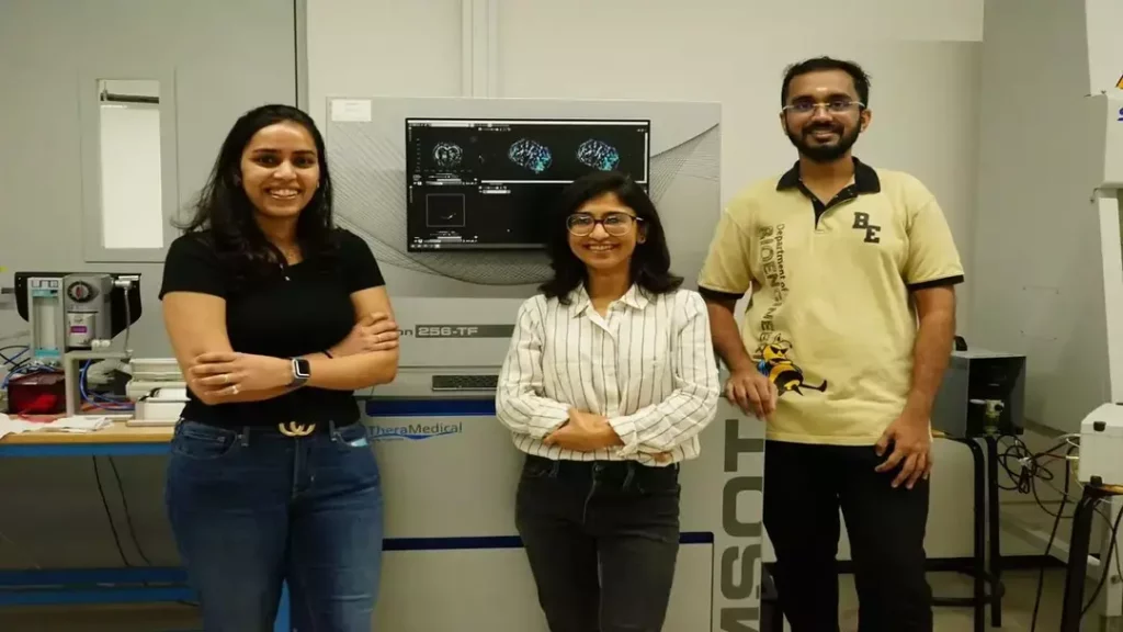
The development of GPc is a significant achievement for the IISc team, led by Dr. Ashutosh Kumar, a professor at the Department of Biological Sciences. Dr. Kumar and his team have been working on the project for several years, and their efforts have finally paid off with the successful creation of a biocompatible imaging molecule that can detect tumours with high accuracy.
“One of the major challenges in developing GPc was finding a molecule that was both biocompatible and effective at detecting tumours,” Dr. Kumar explained in an interview. “We tested numerous molecules before finally finding the right one. Our goal is to make GPc a game-changer in the field of cancer diagnosis.”
So, how does GPc work? The technology uses a combination of light and sound to produce high-contrast 3D images of tumours. Here’s a step-by-step explanation:
- The GPc molecule is injected into the patient’s body, where it accumulates in the tumour tissue.
- A laser is used to shine light onto the tumour, causing the GPc molecule to absorb the energy and convert it into sound waves.
- The sound waves are then detected by a sensor, which translates them into an image of the tumour.
- The image is then reconstructed using a computer algorithm, resulting in a high-contrast 3D image of the tumour.
The benefits of GPc are numerous. For one, it eliminates the need for radiation exposure, which can be a major concern for patients who require frequent imaging. Additionally, GPc is more affordable than traditional PET scans, making it a more accessible option for patients in low-resource settings. The technology also has the potential to be used for imaging other diseases and conditions, such as cardiovascular disease and neurological disorders.
The development of GPc is a significant achievement for the IISc, which has a long history of innovation and research. The institute is home to several renowned research centers and academic departments, including the Department of Biological Sciences, where Dr. Kumar is based.
In an interview, Dr. Kumar expressed his excitement about the potential of GPc to transform cancer diagnosis. “We believe that GPc has the potential to revolutionize cancer diagnosis, particularly in low-resource settings where access to radiation-free imaging technology is limited,” he said. “Our goal is to make GPc a game-changer in the field of cancer diagnosis, and we’re working closely with industry partners to bring this technology to the market.”
The potential of GPc is vast, and its development is a testament to the power of innovation and research. As the technology moves forward, it’s likely to have a significant impact on cancer diagnosis and treatment, particularly in low-resource settings where access to radiation-free imaging technology is limited.